Eukaryotic cells
2.3.1 Draw and label a diagram of the ultrastructure of a liver cell as an example of an animal cell.
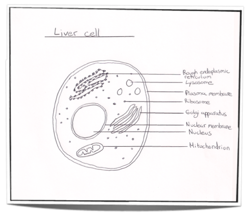
Figure 2.3.1 - Annotated drawing of an animal cell
2.3.2 Annotate the diagram from 2.3.1 with the functions of each named structure.
Ribosomes: Found either floating free in the cytoplasm or attached to the surface of the rough endoplasmic reticulum and in mitochondria and chloroplast. Ribosomes are the site of protein synthesis as they translate messenger RNA to produce proteins.
Rough endoplasmic reticulum: Can modify proteins to alter their function and/or destination. Synthesizes proteins to be excreted from the cell.
Lysosome: Contains many digestive enzymes to hydrolyze macromolecules such as proteins and lipids into their monomers.
Golgi apparatus: Receives proteins from the rough endoplasmic reticulum and may further modify them. It also packages proteins before the protein is sent to it’s final destination which may be intracellular or extracellular.
Mitochondrion: Is responsible for aerobic respiration. Converts chemical energy into ATP using oxygen.
Nucleus: Contains the chromosomes and therefore the hereditary material. It is responsible for controlling the cell.
2.3.3 Identify structures from 2.3.1 in electron micrographs of liver cells.
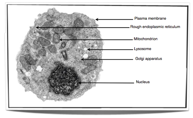
Figure 2.3.2 - Electron micrograph of an animal cell
2.3.4 Compare prokaryotic and eukaryotic cells.
-
Prokaryotic cells have naked DNA which is found in the cytoplasm in a region named the nucleoid. On the other hand, eukaryotes have chromosomes that are made up of DNA and protein. These chromosomes are found in the nucleus enclosed in a nuclear envelope.
-
Prokaryotes do not have any mitochondria whereas eukaryotes do.
-
Prokaryotes have small ribosomes (70S) compared to eukaryotes which have large ribosomes (80S).
-
In prokaryotes there are either no or very few organelles bounded by a single membrane in comparison to eukaryotes which have many of them including the Golgi apparatus and the endoplasmic reticulum.
2.3.5 State three differences between plant and animal cells.
- Animal cells only have a plasma membrane and no cell wall. Whereas plant cells have a plasma membrane and a cell wall.
-
Animal cells do not have chloroplasts whereas plant cells do for the process of photosynthesis.
-
Animal cells store glycogen as their carbohydrate resource whereas plants store starch.
-
Animal cells do not usually contain any vacuoles and if present they are small or temporary. On the other hand plants have a large vacuole that is always present.
-
Animal cells can change shape due to the lack of a cell wall and are usually rounded whereas plant cells have a fixed shape kept by the presence of the cell wall.
2.3.6 Outline two roles of extracellular components.
The plant cell wall gives the cell a lot of strength and prevents it from bursting under high pressure as it is made up of cellulose arranged in groups called microfibrils. It gives the cell its shape, prevents excessive water up take by osmosis and is the reason why the whole plant can hold itself up against gravity.
The animal cell contains glycoproteins in their extracellular matrix which are involved in the support, movement and adhesion of the cell.